Abstract
Potato wart disease is considered one of the most important quarantine pests for cultivated potato and is caused by the obligate biotrophic chytrid fungus Synchytrium endobioticum. This review integrates observations from early potato wart research and recent molecular, genetic, and genomic studies of the pathogen and its host potato. Taxonomy, epidemiology, pathology, and formation of new pathotypes are discussed, and a model for molecular S. endobioticum–potato interaction is proposed.
Taxonomy
Currently classified as kingdom: Fungi, phylum: Chytridiomycota, class: Chytridiomycetes, order: Chytridiales, family: Synchytriaceae, genus: Synchytrium, species: Synchytrium endobioticum, there is strong molecular support for Synchytriaceae to be transferred to the order Synchytriales.
Hosts and disease symptoms
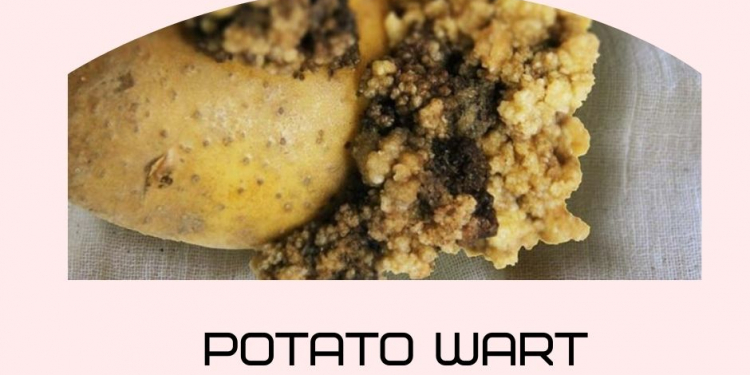
Solanum tuberosum is the main host for S. endobioticum but other solanaceous species have been reported as alternative hosts. It is not known if these alternative hosts play a role in the survival of the pathogen in (borders of) infested fields. Disease symptoms on potato tubers are characterized by the warty cauliflower-like malformations that are the result of cell enlargement and cell multiplication induced by the pathogen. Meristematic tissue on tubers, stolons, eyes, sprouts, and inflorescences can be infected while the potato root system seems to be immune.
Pathotypes
For S. endobioticum over 40 pathotypes, which are defined as groups of isolates with a similar response to a set of differential potato varieties, are described. Pathotypes 1(D1), 2(G1), 6(O1), and 18(T1) are currently regarded to be most widespread. However, with the current differential set other pathogen diversity largely remains undetected.
Pathogen–host interaction
A single effector has been described for S. endobioticum (AvrSen1), which is recognized by the potato Sen1 resistance gene product. This is also the first effector that has been described in Chytridiomycota, showing that in this fungal division resistance also fits the gene-for-gene concept. Although significant progress was made in the last decade in mapping wart disease resistance loci, not all resistances present in potato breeding germplasm could be identified. The use of resistant varieties plays an essential role in disease management.
1 INTRODUCTION
In 1887, Prof. Károly Schilberszky of Budapest University received tubers from two potato fields in Hornyán (now Horňany in the Slovak Republic) of which the entire harvest was affected by wart-like malformations. Initial studies were without success, but in 1896, with new infected tubers, Schilberszky could produce a description of the causal agent of potato wart disease: Synchytrium endobioticum (Schilberszky) Percival (Schilberszky, 1896, 1930). Currently the obligate biotrophic Chytridiomycota (chytrid) fungus is regarded as one of the most important quarantine pests for cultivated potato worldwide (Hampson, 1993; Obidiegwu et al., 2014), and is included on the United States Department of Agriculture (USDA) and Department of Health and Human Services (HHS) Select Agent list (Anonymous, 2018).
Researchers studying fungi at the dawn of plant pathology had to rely on morphological and morphometrical characteristics that could be analysed under a microscope for classification, developmental biology, and the interaction of species with their surroundings. In the case of S. endobioticum, microscopic observations dating back to the 1920s are still among the most accurate descriptions available. In the last years, several research papers appeared that greatly increased our knowledge of the (genome) biology of this pathogen and its interaction with potato. In this review paper we combine knowledge from early S. endobioticum research with recent molecular work and present suggestions for future research to improve disease management.
2 AETIOLOGY AND TAXONOMY
Chytridiomycota, the basal fungal lineage to which S. endobioticum belongs, arose 1000 to 1600 million years ago (Heckman et al., 2001) and is characterized by its motile flagellated spores (zoospores) and an absence of hyphae or mycelium. Chytrids inhabit aquatic and moist terrestrial environments and are free-living saprophytes or pathogens for plants and animals (Barr, 2001; Sparrow, 1960). Initially, S endobioticum was placed in a newly erected genus in the order Chytridiales and was named Chrysophylyctis endobiotica (Schilberszky, 1896). Based on morphological and cytological resemblance to members of the genus Synchytrium, it was later renamed Synchytrium endobioticum (Curtis, 1921; Percival, 1910).
The genus Synchytrium, erected in 1865 with the obligate biotrophic Synchytrium taraxaci pathogen on dandelion as type species, contains approximately 200 species. Previously, all Synchytrium spp. were believed to be parasitic species for algae, mosses, ferns, and flowering plants (Karling, 1964). This view changed with the recent description of the first saprobic member of the genus, that is, Synchytrium microbalum JEL517, which was isolated from an acidic pond in Hancock County, Maine, USA (Longcore et al., 2016). Species within the genus are assigned to one of six subgenera based on variations in the life cycle and morphological features, and S. endobioticum was originally placed in the subgenus , as resting spores were believed to give rise to zoospores directly. However, the work of Kole (1965), which was later confirmed by Sharma and Cammack, Lange and Olson, and Hampson (Hampson, 1986; Kole, 1965; Lange & Olson, 1981c; Sharma & Cammack, 1976), provided evidence that resting spores function as a prosorus, that is, a vesicle in which sporangia are formed. These observations resulted in the transfer of S. endobioticum to the subgenus Microsynchytrium.
Based on light and electron microscopy studies of the zoospore ultrastructure, the placement of the genus Synchytrium in the order Chytridiales is questioned (Barr, 1980; Lange & Olson, 1978a) and the order classification needs revision. Molecular phylogeny of chytrid species based on the rDNA operon (18S, 28S, and 5.8S) (James et al., 2006) showed that the order Chytridiales was polyphyletic, but 18S rDNA sequences could not confirm or refute Synchytrium within the order Chytridiales (Smith et al., 2014). Recent phylogenies based on the mitochondrial and nuclear genomes of representatives of Chytridiomycota strongly support the transfer of the genus Synchytrium to the order Synchytriales (van de Vossenberg et al., 2018, 2019c) (Figure 1). Establishing a robust taxonomical position is an important milestone as it provides the correct evolutionary context. This is particularly relevant for S. endobioticum as it is only distantly related to well-studied plant pathogens in Ascomycota and Basidiomycota, and therefore many features are expected to be distinct or evolved independently from other plant-pathogenic fungi.

3 LIFE CYCLE
Synchytrium species vary considerably in the complexity of their life cycles, and S. endobioticum belongs to the long-cycled species (Karling, 1964). The life cycle of S. endobioticum was described in detail by Curtis (1921), and her observations are largely unchallenged today (Figure 2). During the resting period, while the warted host tissue decays and resting spores are released in the soil, a new inner wall layer is formed inside the resting spore, which acts as a prosorus. Upon germination, the outer spore wall ruptures and the newly formed sorus (also zoosporangium) is released that subsequently gives rise to numerous (200–300) uninucleate haploid zoospores (Lange & Olson, 1981c). ). Released zoospores encyst on the host cell wall within an hour, after which the fungal thallus (or initial cell, sensu Karling, a name used to describe this fungal structure that is distinct from hyphae) penetrates the host cell wall leaving the cyst wall outside the host cell (Weiss, 1925). It is not known if the penetrating fungal thallus is surrounded by the host plasma membrane, similar to haustoria in fungi and oomycetes (Szabo & Bushnell, 2001), or if it is directly in contact with the host cytoplasm. A host cell can be infected by a single or multiple zoospores (Lange & Olson, 1981b). The host nucleus enlarges, becomes irregular in shape, and is often found closely appressed to the developing fungal thallus. 19211921) or summer spores. ). Curtis reported that during this repeated mitosis, five minute chromosomes could be observed (Curtis, 1921). At this stage, brown discolouration of the host tissue can be observed with light microscopy. Hypertrophic growth (cell enlargement) is induced in cells surrounding the infected host cell. Simultaneously, hyperplasia (cell multiplication) increases the amount of meristematic tissue, which enhances the chance of reinfection of the host (Hampson, 1993). Enlarged host cells burst open and release the individual zoosporangia, which in turn release the zoospores, which are morphologically indistinguishable from the zoospores that emerge from resting spores. These zoospores can reinfect the host tissue and start the asexual replication cycle anew. Alternatively, the zoospores may also act as isogametes, fusing to form a diploid biflagellate zygote (sensu Curtis) that can infect the host in a similar fashion as the haploid zoospores (Curtis, 1921). However, instead of forming summer spores, host cells infected with the zygotes give rise to the thick-walled resting spores. The resting spore measures 35–80 μm in diameter and can remain viable and infectious in soils at depths of 50 cm for decades (Przetakiewicz, 2015b). The thick outer wall of the resting spore is composed of a chitin–protein complex and consists of the host’s dead protoplasm and cell wall that is united with the fungal wall to create a firm composite structure (Bal et al., 1981; Lange & Olson, 1981a). Even under favourable conditions, only small percentages (c.10% at maximum) of resting spores are induced to germinate (Kole, 1965). However, these low percentages seem to contradict reported findings in annual reduction of inoculum levels ranging from 70% to 99.5% during cultivation of nonhost species (Baayen et al., 2005). Whereas infections with zoospores induce hypertrophic growth and hyperplasia in the host, this is not the case for host cells infected with zygotes (Karling, 1964).

Curtis described the occurrence of karyogamy after fusion of the isogametes and meiotic divisions before resting spore germination, but this has been a subject of debate (Karling, 1964). It remains unclear if this part of the S. endobioticum life cycle involves a sexual or a parasexual cycle. In both cases, isogametes fuse (plasmogamy) after which the nuclei fuse (karyogamy) to form a diploid nucleus. Where a sexual cycle is highly coordinated and involves meiotic divisions, parasexual cycles produce gametes through mitosis where recombination is limited to chromosome reshuffling as a result of haploidization (Sidhu, 1983). Electron microscopy studies have not been able to provide an ultrastructural description of gamete fusion or meiotic divisions (Lange & Olson, 1981b). Core meiosis genes were identified in S. endobioticum, but due to the patchy presence and absence of some genes and the absence of mating type genes in the comparative genomic analysis of chytrid species (van de Vossenberg et al., 2019c), 3a conclusive answer about the ability of S. endobioticum to undergo a sexual cycle remains elusive, yet crucial to understand population genetics and selection.
S. endobioticum has a strictly obligate biotrophic lifestyle and the prolonged co-evolution with the host could have resulted in loss of essential genes from its genome that results in dependence on the host. Genes in essential pathways, including for GTP synthesis, were found to be absent from the S. endobioticum genome (van de Vossenberg et al., 2019c). Loss of such genes hallmarks the tight relationship between the pathogen and its host and could explain the obligate biotrophic lifestyle.
4 DISEASE SYMPTOMS AND HOST PLANTS
Whereas the host is often not seriously affected by infection with Synchytrium spp., this is not the case for S. endobioticum on its major host potato (Karling, 1964). Disease symptoms on potato tubers are characterized by the warty cauliflower-like malformations that are the result of hypertrophy and cell hyperplasia induced by the pathogen. Meristematic tissue on tubers, stolons, eyes, sprouts, and inflorescences can be infected while the potato root system seems to be insensitive (Karling, 1964; Weiss, 1925).
The warts formed on belowground plant parts are compact protuberances. In contrast, malformations on aboveground affected plant parts seem to follow the morphology of the infected plant organ (Figure . 3). Two main types of symptoms can be differentiated on infected aboveground plant parts, that is, leaf malformation and shoot malformations. Normal development seems to be halted in the case of leaf malformation, whereas it is more or less continued in the case of shoot malformations. Also, aerial root formation was observed after infection of shoots, which could indicate a change in the auxin transport (van de Vossenberg et al., 2019b). Many plant pathogens synthesize plant hormones, toxins, and effectors to manipulate hormonal cross-talk and facilitate infection (Ma & Ma, 2016). The S. endobioticum genes involved in the disruption of the phytohormone system, which leads to the induction of hypertrophic growth and hyperplasia, remain to be identified. First reports on comparative proteomics and transcriptomics between warted and unwarted tissues have appeared (Li et al., 2021; Szajko et al., 2019), but additional research is needed to understand how the pathogen manipulates its host and which pathways and genes are involved.

Hosts that can be infected with S. endobioticum are not restricted to susceptible Solanum tuberosum genotypes. Other solanaceous species such as Solanum lycopersicum (tomato), Solanum dulcamara (bittersweet), Solanum nigrum (black nightshade), Solanum nodiflorum (syn. Solanum americanum, American nightshade), Solanum pseudocapsicum (Jerusalem cherry), Solanum villosum (hairy nightshade), Datura metel var. fastuosa (angel’s trumpet), Nicandra physalodes (apple-of-Peru), and Physalis franchetii (Chinese lantern) have been reported as alternative hosts under experimental conditions (Cotton, 1916; Hampson, 1976). In most of these alternative hosts, very little to no hypertrophy was observed, but newly formed resting spores of S. endobioticum were found to be present in the infected tissue.
Interestingly, the potato root system is not affected by the pathogen, whereas roots are colonized in tomato (Hampson & Haard, 1980). Hampson (1976) tested the reaction of 123 tomato cultivars to S. endobioticum pathotypes 2(G1) and 8(F1) and all of the tested combinations resulted in infection of the host (Hampson, 1976, 1979b). Occurrence of S. endobioticum on wild solanaceous species has been reported from Mexico, but this report has not been confirmed by the Mexican authorities (Obidiegwu et al., 2014).
5 DISTRIBUTION, DISPERSAL, AND DISEASE MANAGEMENT
5.1 Distribution
Currently, S. endobioticum has been reported from all continents where potato is cultivated (EPPO, 2018). Potato wart disease is believed to originate from the Andean region, where it co-evolved with its solanaceous hosts (Przetakiewicz, 2008). The disease was most likely introduced in Europe after the great potato famine (1845–1849) that was caused by Phytophthora infestans, when growers were in need of new seed potatoes (Hampson, 1993). Based on descriptions in nonscientific reports, wart disease was likely to be present as early as 1876 (Gough, 1919). Hampson regarded spread from the United Kingdom to the European mainland, Canada, and (via Ireland) South Africa and New Zealand the most likely scenario (Hampson, 1993). In the decades after description of the pathogen, disease reports came in from different countries describing the first occurrence of potato wart disease on their territory, for example, Germany (1908), Ireland (1908), Canada (1909), Sweden (1912), Netherlands (1915), Czech Republic (1915), and Poland (1917) (Baayen et al., 2006; EPPO, 2018; Gough, 1919; Hampson, 1993; Obidiegwu et al., 2014). In 1952, potato wart disease was introduced in India from Danish seed potatoes (Phadtare et al., 1990).
Both world wars played a role in the spread of the disease across the European continent. At the end of the First World War, there was a shortage of ware and seed potatoes, and potatoes from infected areas in the UK were used as seed in districts that were untouched by the disease (Gough, 1919). Similarly, potato wart disease is believed to be introduced in the former Union of Soviet Socialist Republic (USSR) with the movement of German troops at the onset of the Second World War (Hampson, 1979a). After the Second World War, trade with infected seed potatoes has often been suggested as the cause of spread to other countries. More recently, the disease was reported for the first time in Turkey (2003) (Çakır, 2005), Bulgaria (2004) (Dimitrova et al., 2011), and Georgia (2009) (Gorgiladze et al., 2014). Also, recently the disease reoccurred in Denmark (2014), where it was believed to be eradicated since 1989 (Anonymous, 2014).
5.2 Disease management
Countries worldwide implemented control systems to prevent the introduction and spread of S. endobioticum in national or regional legislation. Guidelines for national regulatory control systems for the monitoring, containment, and suppression of S. endobioticum are provided by the European and Mediterranean Plant Protection Organization (EPPO) in standard PM9/005, which describes (a) elements of the monitoring programme that should be conducted to detect a new infestation or to delimit an infested area; (b) measures aiming to suppress the pest where it is found; and (c) measures to contain the pest to prevent further spread in a country or to neighbouring countries (EPPO, 2017b). In short, fields in which the pathogen is found are placed under official control (i.e., “scheduled”). No potato production or the production of plants for planting is allowed for a period of 20 years. A safety zone, or buffer zone, is established and cultivation of only resistant potato varieties is allowed in these areas (EPPO, 2017a, 2017b). A plot can be partially descheduled after a shorter period (at least 10 years) so that ware potato varieties resistant to the pathotype(s) present in the infested plot may be grown (EPPO, 2017a).
To reduce or eradicate the pathogen from infested soils, an impressive number of over 600 different treatments, including application of more than 120 control agents, have been tested up to the 1960s (Hampson, 1988). These treatments were unsuccessful in most cases, and if they did have success to eradicate the pathogen, the control agents used were so toxic that they rendered treated soils barren. The use of crushed crab shell was shown to suppress wart disease in infested fields (Hampson & Coombes, 1995), and crop rotation and intercropping potato with other crops like maize were reported to reduce the number of viable resting spores in soils (Obidiegwu et al., 2014). Research of chemical treatments was largely abandoned from the 1960s as phytosanitary measures and the use of resistant varieties had proven to effectively minimize the spread of the disease.
Cotton (1916) hypothesized that alternative hosts, which are frequently found within and bordering potato fields, could act as reservoirs of the disease and thus promote the survival of the pathogen when only resistant potato varieties are cultivated (Cotton, 1916; Hampson, 1979b). The role of the alternative hosts to act as reservoirs for S. endobioticum and their potential to extend the longevity of the pathogen under field conditions have not been studied and it is recommended to fill this knowledge gap. It was observed though that the presence of nonhost species, Solanum nigrum, and resistant potato varieties on infested plots do not result in a build-up of pathogen inoculum (Baayen et al., 2005).
5.3 Dispersal and tracking pathogen spread
Although the pathogen typically remains restricted to the infested parts of fields, local and long-distance dispersal are encountered. Spread of the disease by human activity was recognized early on, and the movement of infected seed potatoes, soil, and manure were regarded to have the biggest contribution to local and long-distance dissemination of the pathogen (Gough, 1919). Zoospores do not play a role in dissemination over long distances as they were reported to actively migrate over only small distances of up to 5 cm (Franc, 2007). Moreover, zoospores are short-lived, which would further restrict their role in long-range dispersal. In contrast, resting spores have been reported to be transported by water currents (Weiss, 1925) and wind (Hampson, 1996; Jösting, 1909), accounting for dispersal over larger distances. Floor areas of cars, vans, and trucks being operated in Newfoundland (Canada), where the pathogen is widespread, were demonstrated to harbour infectious resting spores (Hampson & Wood, 1997; Hampson et al., 1996). Another vector for long-distance transport is resting spores in inconspicuous warts, when such tubers are used as seeds or industrially processed (Hampson, 1993). Finally, organic waste from processing has to be sanitized before it can be used as fertilizer in agriculture, in horticulture, or in private gardens, for instance by composting or pasteurization (EPPO, 2008). While these techniques have been proven effective against plant pests such as potato cyst nematodes, they have little to no effect on the resting sporangia of S. endobioticum (Steinmöller et al., 2012). Moreover, composting of infected potato material is part of the inoculum preparation for Spieckermann assays (EPPO, 2004). The reliability and reproducibility of pathogen detection in materials and soils has increased with the description of real-time PCR tests based on ITS1, ITS2, and the 18S small ribosomal subunit, which offer increased sensitivity for detection of the spores in soils and other matrices (van Gent-Pelzer et al., 2010; Smith et al., 2009). Additional molecular assays that would allow the specific detection of viable resting spores would further enhance the application of molecular methods. Early pathogen detection combined with track-and-tracing using, for instance, mitogenomic diversity will allow regulatory bodies to take risk-based measures and to identify potential origins and identify or disqualify potential outbreak sources. Epidemiological models can be employed in risk assessment and prediction of future pathogen spread to prioritize regions for phytosanitary surveys, as was demonstrated in a case study for the republic of Georgia (Andersen Onofre et al., 2021).
6 MOLECULAR PATHOGEN–HOST INTERACTION
The genomes of plants and the microorganisms aiming to colonize them are shaped by millions of years of co-evolution (Carella et al., 2018). A model of the molecular S. endobioticum–potato interaction (Figure 4) can be sketched by combining insights based on light microscopy and electron microscopy studies on the interaction (Cartwright, 1926; Curtis, 1921; Hampson, 1986; Lange & Olson, 1978b, 1981a, 1981b, 1981c), general knowledge of fungal plant–pathogen interactions (reviewed in Carella et al., 2018; Lo Presti et al., 2015; Selin et al., 2016; de Wit et al., 2009), and recent genomic studies (van de Vossenberg et al., 2019a, 2019c).

6.1 Invading the host
After being released from either summer spores or resting spores, zoospores of S. endobioticum have to identify and move towards a suitable host cell, on which they encyst before penetration. Zoospores of plant-pathogenic fungi and oomycetes are known to direct their movement by responding to external stimuli (Islam & Tahara, 2001). In their review, Obidiegwu et al. (2014) mention the research performed by Esmarch (1924–1928) on the response of S. endobioticum zoospores to chemotactic stimuli of the host. Unfortunately, we could not retrieve additional information about the nature and effects of these stimuli. From the analysis of the genomes of the pathogenic chytrids S. endobioticum and Batrachochytrium dendrobatidis, expansions of genes associated with chemotaxis were observed relative to saprobic chytrid species. In addition, genes involved in the G-protein-coupled receptor signalling pathway were found enriched in S. endobioticum relative to nine other chytrid species (van de Vossenberg et al., 2019c). Membrane-bound G-protein-coupled receptors sense environmental signals (Xue et al., 2008), and in the case of S. endobioticum they could be involved in sensing specific host-derived signals.
When attempting to encyst on the host cell before penetration, the invading zoospore has to evade and/or suppress the plant innate immune system. Plant membrane-bound pattern recognition receptors (PRRs) trigger the immune system upon recognition of conserved pathogen-associated molecular patterns (PAMPs). The defence response triggered by PRRs is called PAMP-triggered immunity (PTI), and leads to chemical and physical adjustments that arrest pathogen proliferation. Such responses include the production of reactive oxygen species, rapid changes in hormone biosynthesis, and cell wall reinforcement by callose deposition (Toruño et al., 2016). PTI is considered to be effective against nonadapted microbes, and is generally regarded as a first layer of the plant defence system (Lo Presti et al., 2015; Zipfel, 2008). Breakdown products of chitin, an insoluble cell wall component of fungi also present in S. endobioticum, act as PAMPs. The fungus needs to secrete effector proteins that either mask the chitin oligomers or suppress the subsequent host immune response. In plants, several membrane-bound chitin receptors have been identified that all contain extracellular LysM domains (Kombrink et al., 2011). In fungi, LysM effectors have been reported that interfere by binding to chitin and thereby prevent chitin-triggered immune responses (Bolton et al., 2008). In S. endobioticum, LysM-containing proteins were found enriched relative to all nine nonplant–pathogenic chytrid species analysed (van de Vossenberg et al., 2019c). These LysM-containing proteins could have a function in the interference of chitin-triggered immunity or the protection against chitinases, or act as killer toxins to combat competing fungi (Tzelepis & Karlsson, 2019).
The plant innate immune system is not only triggered by conserved PAMPs. Several plant cell wall degradation products are categorized as damage-associated molecular patterns (DAMPs) and can lead to DAMP-triggered immunity (DTI) (Gust et al., 2017). Reduced cell wall-degrading enzyme (CWDE) content was previously found in the stealth-like pathogen Zymoseptoria tritici (Goodwin et al., 2011) and the biotrophic and symbiotic fungus Lacaria bicolor (Martin et al., 2008). In these plant–microbe systems, it was hypothesized that a reduced CWDE repertoire may prevent DTI (Choi & Klessig, 2016). Relative to its saprobic sister species S. microbalum, S. endobioticum has a reduced set of CWDEs, and it is likely that S. endobioticum employs a stealth-like strategy to evade detection and establish its biotrophic lifestyle (van de Vossenberg et al., 2019c).
6.2 Inside the host cytoplasm
Currently it is not known how effectors reach the host cytoplasm. More study is needed to determine if an uptake mechanism exists, as has been described for other fungal and oomycete pathogens. But after penetration of the host, the fungal thallus can directly secrete effector proteins into the host cell to manipulate the metabolism, which results in hypertrophy and hyperplasia of potato tissue. In contrast to the highly conserved PAMPs and microbe-associated molecular patterns, effectors are very diverse, often specialized towards a specific host, and species- or even strain-specific. The complex effector repertoire of plant-pathogenic fungi is believed to influence the fungal lifestyle and host specialization (Lo Presti et al., 2015). Plants have responded to effector-induced suppression of PTI by evolving highly specific resistance (R) proteins. These intracellular receptors belong to the conserved nucleotide-binding and leucine-rich repeat domain-containing (NLR) superfamily, and can interact directly with pathogen effectors or indirectly involving effector targets or structural mimics of such targets (Jones et al., 2016; Lo Presti et al., 2015). Upon recognition of effector proteins by the cognate R proteins, the second layer of the plant defence system, called effector-triggered immunity (ETI), is activated. Effects of ETI are the induction of pathogenesis-related genes and a local hypersensitive response (HR), which is visible as localized cell death at the site of infection. Effectors that are recognized by R proteins are referred to as avirulence (AVR) proteins. For S. endobioticum, only a single effector has been described so far, AvrSen1 (van de Vossenberg et al., 2019a), which matches the potato Sen1 R gene. This is also the first effector that has been described in Chytridiomycota, which shows that in this division of fungi resistance also fits the gene-for-gene concept. Microscopic observations showed that zoospores penetrate resistant varieties and susceptible plants equally well (Cartwright, 1926), so no differentiation between different pathotypes is expected in the effector repertoire used to suppress PAMP-triggered immunity in the extracellular phase. However, in an incompatible interaction (e.g., AvrSen1–Sen1) the pathogen dies within a few hours after penetration of the host as a result of what has been described as pathogen-induced defence necrosis (Cartwright, 1926; Karling, 1964). The AvrSen1 effector is expected to be recognized intracellularly, as it triggers an HR upon expression in the plant cytoplasm. This corresponds with the intracellular localization of the presumed Sen1 gene product, which has homology with TNL receptors (Prodhomme, 2020).
6.3 Effector evolution
As ETI is a key mechanism for wart disease resistance, it requires knowledge of effector composition and distribution in S endobioticum populations, illustrated by the cloning of AvrSen1. Pathotype 1(D1) isolates are defined by their avirulence on Sen1 plants. This distinction was used to identify AvrSen1, which was presumed to be present in pathotype 1(D1) isolates but (functionally) absent in “higher pathotypes” (see section 7). Five different AvrSen1 variants were found in higher pathotypes that could not trigger HR in Sen1 plants. This showed that AvrSen1 mutated multiple times during evolution, presumably to evade detection by Sen1 (van de Vossenberg et al., 2019a). In several higher pathotype populations, which overcome Sen1 resistance, the functional AvrSen1 was also found at very low frequencies beside the variants (van de Vossenberg et al., 2019a). Apparently, AvrSen1 is maintained at low levels in the virulent fungal population, which could suggest a balanced selection in which AvrSen1 has a function in virulence. However, such a proposed virulence function is not essential in repeated cycles of propagation on Sen1 plants through summer spores (i.e., Glynne–Lemmerzahl assay), which caused AvrSen1 to become undetectable in the population. As for many other fungal effectors (Lo Presti et al., 2015), AvrSen1 lacks identified functional domains or a predicted function. The absence of nuclear, mitochondrial, or chloroplast localization signals suggests a cytoplasmic target. In the Canadian pathotype 6(O1) isolate LEV6574, the truncated AvrSen1 gene is expressed in vivo (van de Vossenberg et al., 2019c). The function and the importance of AvrSen1 during infection are not yet known. Even though fungal effectors are often considered to be dispensable due to functional redundancy, a trade-off between the need to maintain an effector as it has a role in virulence and the need to avoid recognition by the host plant is likely to exist (Sharpee & Dean, 2016). Whether the truncated proteins can act as functional homologues and if other functional homologues are present in higher pathotypes are questions that remain to be addressed.
6.4 Breeding novel resistant potato varieties
Potato varieties resistant to potato wart disease were reported as early as 1909, and by 1919 immunity trials performed in the UK, Germany, and the USA resulted in the publication of a list with 64 potato varieties that were resistant to the disease (Gough, 1919). Interestingly, potato wart disease resistance was one of the first traits in plants studied for Mendelian inheritance (Salaman & Lesley, 1923). Classical breeding for resistance began around a century ago and was very successful in selecting varieties resistant to the disease. The use of resistant varieties and strict phytosanitary measures were so successful that breeding for potato wart resistance lost priority until the discovery of new pathotypes that caused disease in previously resistant varieties (Obidiegwu et al., 2015). This resistance breakdown distinguished pathotype 1(D1) from the higher pathotypes. Several resources for wart disease resistance could be distinguished, and quantitative resistance locus (QRL) analyses identified genomic regions providing resistance to (combinations of) the pathotypes of major importance on every chromosome (Obidiegwu et al., 2015; Prodhomme et al., 2020a). In these mapping studies, major- and minor-effect QRLs could be distinguished. A distinct naming system for major- and minor-effect QRLs was therefore established. The major QRLs are referred to as Sen genes (from S endobioticum), followed by a sequential number.
The major R gene responsible for pathotype 1(D1) resistance was named Sen1 and Hehl et al. mapped it to the distal end of the upper arm of chromosome 11 (Hehl et al., 1999). Further fine-mapping and functional analyses showed that Sen1 encodes a TNL-type immune receptor (Prodhomme et al., 2020a). Sen1 gene is widespread in potato breeding germplasm (Prodhomme et al., 2020a, 2020b) and was already present in old varieties such as Pink Fir Apple (1850), Champion (1876), and Belle de Fontenay (1885), which suggests Sen1 was present in the European breeding material before the first introduction of S. endobioticum in Europe (Prodhomme et al., 2020b). Sen1, although being the major pathotype 1(D1) resistance source, does not explain all the pathotype 1(D1) resistance present in the potato breeding material. A second independent dominant gene for pathotype 1(D1) resistance (Sen1-4), which genetically mapped to the long arm of chromosome 4, was identified by Brugmans et al. (2006). The distribution of this gene in breeding material was not studied elaborately and it is unknown if it is an important source of resistance to pathotype 1(D1) or any other pathotype.
Resistance to the higher pathotypes is observed less frequently in potato-breeding germplasm and the identification of loci providing resistance to one or several of these pathotypes is crucial to develop new resistant varieties. Sen2 provides broad resistance to pathotypes 1(D1), 2(Ch1), 2(G1), 3(M1), 6(O1), 8(F1), 18(T1), and 39(P1) and has been mapped on chromosome 11, approximately 32 Mb distant from the Sen1 locus (Plich et al., 2018). Sen2 was identified in a complex diploid potato hybrid with multiple wild species origins and is probably not yet introgressed in commercial varieties (Prodhomme et al., 2020a). Sen3 also provides broad resistance to pathotypes 1(D1), 2(G1), 6(O1), and 18(T1) and resides at the same locus as Sen1, on the upper arm of chromosome 11 (Bartkiewicz et al., 2018; Obidiegwu et al., 2015; Prodhomme et al., 2019). This gene is mainly found in German and Polish potato varieties descending from the potato clone BRA9089, the putative unique donor of Sen3 in commercial varieties (Prodhomme et al., 2019, 2020a). Sen4 provides resistance to pathotypes 2(G1), 6(O1), and 18(T1) and has been mapped to the long arm of chromosome 12. Sen4 is mainly found in Dutch starch varieties that descend from the breeding clone AM78-3704. In addition, Sen4 is found in the German variety Panda but probably from a different source than the Dutch material (Prodhomme et al., 2020a). Currently, Sen5 is very rare in the potato breeding material, having been identified in only three commercial Dutch starch cultivars so far. Sen5 can be traced back to the common ancestor VE71-105 and was probably introgressed in breeding germplasm from Solanum vernei (Prodhomme et al., 2020a).
Although significant progress was made in the last decade in mapping wart disease resistance loci, not all resistances present in potato breeding germplasm could be identified. This is the case even for varieties in the EPPO differential set. For instance, the strong resistance to pathotypes 2(G1), 6(O1), 8(F1), and 18(T1) observed in the variety Belita probably originates from the wild species S. vernei. This resistance is distinct from Sen5, as Belita is negative for the Sen5 markers. The resistance from Belita remains to be characterized genetically. Also in the EPPO set, the resistance observed in the variety Saphir to the pathotypes 6(O1), 8(F1), and 18(T1), which may have been inherited from the breeding clone MPI 44.1016/24, remains to be identified. Other potato varieties negative for all the Sen genes described so far but resistant to one or several pathotypes have been reported (Prodhomme et al., 2020a). These indicate that additional resistance sources already present in the commercial gene pool can be further characterized and exploited. Interestingly, the mapping of both major- and minor-effect QRLs indicated that they colocalize with clusters of receptors involved in innate immunity (Hehl et al., 1999). ETI is therefore probably the main determinant for wart disease resistance. ETI is easily overcome when a single effector is lost in the pathogen and it is generally acknowledged that the stacking of multiple ETI receptors in crop varieties is mandatory to enhance durability of resistance (Luo et al., 2021).
In the process of selection of effective wart disease resistance in breeding programmes, major- and minor-effect QRLs have been stacked, indicating this may be a requirement for the effective control of the pathogen (Prodhomme et al., 2020a). Varieties with only one R gene may show incomplete resistance, especially to the higher pathotypes whose isolates are genetically very complex. In order to enhance the durability of wart resistance in novel varieties, stacking of wart disease QRLs might therefore be an important aspect of breeding strategies. In such stacking schemes, the use of Sen genes that recognize a broad spectrum of isolates is preferred in order to reduce the number of loci to be combined.
7 PATHOTYPING SYSTEMS
In 1931, Köhler stated that the pathogen had a uniform make-up and differences between isolates of the pathogen were not known (Hampson, 1993). However, in the 1930s, resistant potato varieties from the UK grown in Newfoundland (Canada) were found to be very susceptible to the local strain of S. endobioticum (Olsen, 1961), and in the decades afterwards the existence of different S. endobioticum pathotypes became clear. For S. endobioticum over 40 pathotypes are currently recognized, which are defined as groups of isolates with a similar response to a set of differential potato varieties. After the Second World War, when the European continent was politically split in an eastern and western part, different naming conventions were implemented to identify the new infection outbreaks. In eastern Europe new pathotypes were named after the origin where they were found, but in western Europe sequential Arabic numbers were used to define new pathotypes. As a consequence, the first new officially described pathotypes found in Gießübel and Silberhütte in 1941 were named G1 and SB in the east but 2 and 3 in the west, respectively. Baayen et al. (2006) proposed a harmonization of the pathotype naming system in 2006, which is still used today (Baayen et al., 2006). Under this system, both naming conventions are combined, for instance 2(G1) for the pathotype 2 isolate identified in Gießübel. In the years following the description of pathotypes 2(G1) and 3(SB), new S. endobioticum pathotypes were reported from Germany, Poland, the Czech Republic, and the Ukraine, based on their interaction with differential panels of locally popular resistant and susceptible potato varieties. Among these new pathotypes are the German pathotypes 6(O1) (found in Olpe, 1952), 8(F1) (Fulda, 1954), and 18(T1) (Trannroda, 1978) (Baayen et al., 2006). The initial S. endobioticum pathotype is nowadays known as pathotype 1(D1), and collectively nonpathotype 1(D1) isolates are referred to as higher pathotypes. However, it is important to note that this does not imply that higher pathotypes descended from pathotype 1(D1). Pathotypes 1(D1), 2(G1), 6(O1), and 18(T1) are currently regarded to be most widespread and of major importance. The latest two formally described pathotypes are 38(Nevşehir) from Nevşehir (Turkey, 2009) and 39(P1) from Piekielnik (Poland, 2015) (Çakır et al., 2009; Przetakiewicz, 2015a). Although not published in a peer-reviewed journal, pathotypes 40(BN1) and 41(P2) have been reported from Poland (Anonymous, 2015) and Sweden (Björklund & Boberg, 2018).
Along the years, many different and locally preferred differentially resistant varieties were used for pathotyping. It was only in the beginning of the 21st century that the EPPO published a harmonized differential set of potato varieties that could be used as a diagnostic standard for S. endobioticum isolates (EPPO, 2004). For reasons of simplicity and reproducibility, the EPPO differential set was rather small (Baayen et al., 2006; Flath et al., 2014). It was selected for its ability to distinguish the pathotypes of major agricultural importance. Two main methods are used to assess the interaction between host and pathogen: Spieckermann (Spieckermann, 1924) and Glynne–Lemmerzahl (Glynne, 1925; Lemmerzahl, 1930); both are based on the inoculation of potato tubers. In the Spieckermann assay, the inoculum consists of resting spores, which are the result of a (para)sexual cycle. In contrast, fresh warts containing summer sporangia, which are produced asexually, are used as inoculum in the Glynne–Lemmerzahl assay. Recently, a new infection procedure was described based on infection of aboveground plant parts or auxiliary buds demonstrating that R genes are expressed in both aetiolated belowground sprouts and green aboveground organs (van de Vossenberg et al., 2019b). Although the methods are different, the resulting scores are mostly well correlated although sometimes consistent differences appear to exist (Prodhomme et al., 2020b).
7.1 Intraisolate diversity
S. endobioticum isolates represent communities in which multiple genotypes coexist. This is not only true for mitochondrial genotypes (Figure S1), but also for the nuclear-encoded species-specific secretome and AvrSen1 gene variants (van de Vossenberg et al., 2018, 2019a). Particularly striking is the AvrSen1 variation found within isolates of higher pathotypes. These isolates are no longer recognized by the Sen1 gene, resulting in a compatible interaction and proliferation of the pathogen. However, the avirulent form of the gene was still found to be present at low frequencies in isolates that were multiplied on the potato variety Deodara, which is susceptible to all S. endobioticum pathotypes. Conversely, in pathotype 1(D1) isolates carrying the avirulent AvrSen1 gene, virulent forms of the gene (i.e., loss of AvrSen1) were present (van de Vossenberg et al., 2019a). This was not directly apparent from genomic sequence data, but two multiplications of a pathotype 1(D1) isolate on the semiresistant potato variety Erika resulted in a pathotype 6(O1) phenotype (van de Vossenberg et al., 2018). This increased virulence is possibly a result of selecting against S. endobioticum genotypes carrying the dominant avirulence gene. Near-complete loss of AvrSen1 in the isolate producing the 6(O1) phenotype was verified by amplification of the AvrSen1 gene followed by PacBio SMRT sequencing (van de Vossenberg et al., 2019a). Multiplications leading to the observed selective sweep were performed with the Glynne–Lemmerzahl bioassay, in which asexually produced zoospores from fresh warts are used as inoculum, suggesting that the diversity was already present within the initial inoculum and did not require recombination in a (para)sexual cycle.
7.2 An advanced pathotyping system
Differential sets are indispensable for the identification of the R genes that recognize the genotypes in a defined pathogen population. New genotyping methods provide superior resolution of pathogen population structures and, likewise, new genotyping methods and pedigree information of potato varieties allow the identification of specific haplotypes carrying R genes. To facilitate the implementation of cultivar resistance in pathotype identification and disease management, it is important to establish a more transparent link between the potato varieties used in the differential set and the commercially used potato varieties. A catalogue of S. endobioticum R genes in the potato breeding pool including the EPPO differential set was recently described (Prodhomme et al., 2020a). The S. endobioticum R gene content for 141 commercial potato varieties was assessed, and it was shown that the R genes Sen2, Sen4, and Sen5 are not represented in the EPPO differential set. Moreover, Sen1 and Sen3 are present in multiple combinations with other unknown R genes (Prodhomme et al., 2020a). To improve the predictive value of the differential set for resistant cultivar selection and pathotype identification, novel potato varieties need to be selected for the EPPO differential set that harbour defined single combinations of R genes and comprise the complete set of known and deployed Sen R genes. A similar optimized differential set for late blight resistance was produced using a combination of molecular markers and effector responses (Zhu et al., 2015). A major limitation for making reliable differential sets in potato is that the R genes will be present in different genetic backgrounds, when produced through classical tetraploid breeding. In such diverse genetic backgrounds, there might be different genes present that positively affect the spectrum of resistance, including minor QRLs recognizing minor AVR proteins in the pathogen population. Conversely, other genes could negatively affect the activity of the R gene. Ideally, a differential set is generated in an isogenic background. This can be achieved using transformation of cloned R genes into one variety (Zhu et al., 2015), or it could be achieved through inbred lines (Su et al., 2020). As none of the Sen R genes is currently cloned and inbred lines are difficult to generate, this will probably remain a concept for the coming decade. In addition, it is important to establish a set of S. endobioticum reference material that represents the genetic diversity that is present at different geographic locations and further characterized based on the mitochondrial haplotype, SSR profile, molecular characterization, and avirulence profile.
7.3 Molecular classification as track-and-trace tool
As the EPPO differential set of potato varieties was designed to distinguish isolates of major agricultural importance, other pathogen diversity remains undetected (van de Vossenberg et al., 2018). This became even more prominent when a study of the European population demonstrated that multiple S. endobioticum introduction events have occurred, reflected by their distinct mitochondrial genome sequences. Notably, pathotypes 2(G1) and 6(O1) occur in clearly distinct genetic backgrounds. As pathotype profiles are phenotypes that can be shared by multiple distinct genotypes, they represent an inherently inaccurate classification system (van de Vossenberg et al., 2018). Moreover, the molecular clustering of isolates seems to be more consistent because a similar clustering was observed with mitochondrial sequences, nuclear secretome sequences, and microsatellite loci (Busse et al., 2017; Gagnon et al., 2016; van de Vossenberg et al., 2019a). Therefore, a pathotype classification system must be based on more data than just the phenotypes on the small differential set of potato varieties. Similarly, isolates from a specific location were found to harbour multiple genotypes. It is envisaged that genomic studies in combination with traditional pathotyping using an updated differential set of potato varieties will provide a pathotyping system that is of higher value for agricultural practice. Although such a functional pathotyping system still requires technical limitations to be overcome, good progress has been made in recent years. For instance, the genomic characterization of isolates is still challenging given the difficulties to obtain material that is pure enough for whole genome sequencing and molecular pathotyping. Also, the number of known fungal effectors that match the potato R genes is still limited. Currently it is only possible for AvrSen1, that is, the fungal effector pathotype recognized by Sen1 plants resulting in pathotype 1(D1) resistance (van de Vossenberg et al., 2019a). This could be extended when additional effectors or other functional determinants become available. Also, an improved pangenomic approach in which the genome is dissected into conserved and accessory parts could contribute to novel pathotyping tools. The core can be exploited for track-and-trace studies, while the accessory part can be studied for functional diversity. Although still in its infancy, the growing number of genomes that are being currently generated could provide a window of opportunity for such pangenome analyses.
8 CONCLUDING REMARKS
S. endobioticum, the causal agent of potato wart disease, is an important pathogen with a worldwide quarantine status that is included on the USDA and HHS Select Agent list. The pathogen keeps re-emerging as a threat to potato production as shown by the notification of outbreaks in recent years in different regions in the world. The obligate biotrophic lifestyle, the distinct taxonomic position, and the quarantine status itself hamper research. Nevertheless, the pathogen and the disease have been studied for more than a century, and in the last decade considerable progress has been made in our understanding of S. endobioticum and the interaction with its host potato. The application of genomics has contributed significantly to these advances and allowed the assessment of the S. endobioticum gene content and its phylogenetic position and the identification of the first AVR gene. Furthermore, it has opened up opportunities to track-and-trace the pathogen outbreaks using molecular assays or sequence analysis. This has generated new insights in the diversity and the pathology of S. endobioticum. Still, many aspects of the pathogen remain unknown, including parts of the life cycle and the pathogen–host interactions. In particularly, linking genome information with genetics could provide more information on the presence of a (para)sexual cycle in S. endobioticum and consequently on the potential of recombination and selection and on the diversity within isolates. On the host side, the molecular characterization of resistance in potato allowed the identification of genomic regions in which resistance loci reside. Further characterization of resistance loci, including the cloning of the first Sen R gene, would be an important milestone to study the interaction on a molecular level. Human activity including transport of equipment and soil and trade of produce and plant debris, flooding, and wind erosion are important means by which the pathogen can spread. With increased global food demands, the need for circular agriculture, and climatic changes, these events are likely to increase. New tools and optimized disease management based on monitoring and the use of resistant potato varieties are needed to control potato wart disease in future decades.
ACKNOWLEDGEMENTS
The authors would like to thank NPPO-NL staff members Johan Meffert for providing images of S. endobioticum resting spores and Sietse van der Linde for reviewing this manuscript prior to submission.


